Overview
The primary research interests in the Tissue Biomechanics Laboratory relate to the biomechanical function, degeneration and regeneration of cartilaginous tissues, including temporomandibular joint (TMJ) cartilage, intervertebral disc (IVD), articular cartilage, and cornea. The research is conducted at multiscale levels from whole body to single cells, including imaging based joint kinematics (body level), tissue characterization and constitutive modeling (tissue level), single cell mechanics and mechanobiology (cell level), and molecular transport and assembling (molecular level). Each hierarchical scale has specific advantages and disadvantages, so the ability to cross different levels of study, as well as different in vitro and in vivo model systems, has proved enormously valuable in our ability to translate findings from one system to another. This approach provides new perspectives on our understanding of cartilaginous tissue degeneration and regeneration processes by bringing together the principles of biomechanics and biology.
Current research areas include the biomechanical mechanisms of cell mediated tissue degeneration, novel imaging techniques for nondestructive assessment of cartilage composition and structure, and interactions between mechanical, electrical, chemical and matrix-supplied cues in controlling the development of functional engineered tissues. Our work has contributed to the world's first demonstration of a functional full synovial joint regeneration (Lancet, 2010).
1. Temporomandibular Joint Biomechanics and Mechanobiology
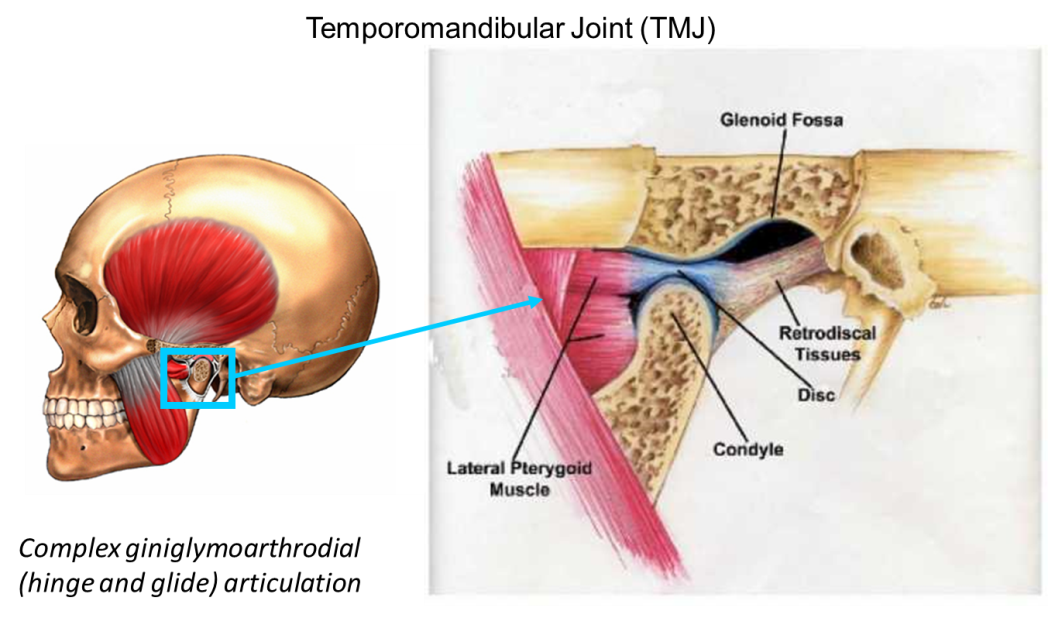
Temporomandibular joint disorders (TMJD) affect approximately 35 million people in the US with tremendous morbidity and financial cost, yet its etiology remains poorly understood. In approximately 30% of TMJD patients, mechanical dysfunction of TMJ disc, especially displacement due to tissue degeneration, is a common event. Degenerative changes generally occur more than a decade earlier in the TMJ than other human joints (e.g., knee and hip) for unknown reasons. TMJD surgical intervention is often unsuccessful, thus, research to understand the pathophysiology of TMJ disc degeneration for earlier diagnosis and management are essential. The TMJ is the most structurally and mechanically complicated load bearing joint in the body. It is generally believed that pathological mechanical loadings, e.g. sustained jaw clenching or malocclusion, trigger a cascade of molecular events leading to TMJ disc degeneration. Mechanical loading on TMJ disc can result in complex physicochemical environment changes surrounding disc cells. The magnitude/frequency of these physicochemical signals and the specific cell responses to the signals determine the path of normal remodeling or degeneration. To date, the quantitative relationships between the loading event (body level), signals (tissue level), and cell response (cell/molecular level) are unavailable in any joints. The long-term goal of this project is to establish such quantitative relationships and use them to develop a model both elucidating the biomechanical etiology of disc degeneration as well as aid in identifying bio-indicators for earlier diagnosis of TMJD. We hypothesize that the nutrient concentrations and cell metabolic rates in TMJ disc are sensitive to the pattern and magnitude of the mechanical loading during jaw function and are therefore potential early bio-indicators for evaluating the impact of mechanical loading on TMJD.
TMJ function and kinematics (body level). Develop techniques to combine three-dimensional TMJ anatomies and real-time kinematics to permit a subject specific dynamic analysis of the intra-articular space. The goal is to conduct patient specific TMJ dynamic functional assessment, which will provide individual TMJ disc deformation and motion during various oral function and TMJ loading.
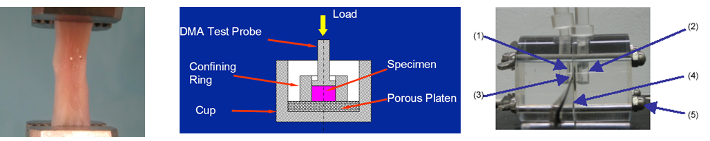
TMJ tissue biomechanical and biochemical characterization (tissue level). Develop tensile, compression, and shear testing techniques to characterize the viscoelastic mechanical properties of TMJ cartilage tissues. Develop techniques, such as electrical conductivity device and diffusion cell, to determine ion and nutrient diffusivities. The goal is to establish constitutive relationships between the mechanical/transport properties and tissue composition and structure.
Effect of physicochemical environment on cell homeostasis (cell level). Study the effect of physical and chemical environments on viability, volume regulation, metabolic state, and matrix synthesis of TMJ cartilage cells. The goal is to elucidate the mechanism of mechanotransduction to integrate biomechanics and cell biology in order to understand TMJ pathophysiology.
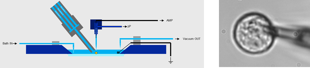
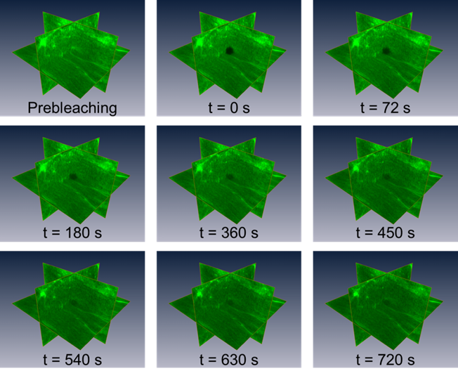
3D anisotropic molecular mobility and assembling (molecular level). Develop novel techniques, such 3D FRAP, to investigate signaling molecules and matrix protein transport and assembly in cartilaginous tissues.
TMJ finite element model (multiscale). The multiscale finite element model, integrating joint imaging and kinematics, tissue mechanics, and cell metabolism, will produce data thatcharacterize the effect of dynamic contact mechanics on cell behavior as a contributing factor to the tissue degeneration. Such data have never before been collected from living humans. The outcomes of this project will closely address the current clinical barriers for treating TMJ disorders, i.e., inadequacy of current understanding of the causes and the lack of early diagnostic tools. More broadly, the proposed approach may serve as a template in understanding the earliest development of articular tissue degeneration in the tibiofemoral (knee) joint, which affects 30% of older North Americans. This project will also provide foundational transport and energy metabolic data for TMJ tissue regeneration since nutrition is a key prerequisite for cartilaginous tissue engineering.
2. Intervertebral Disc
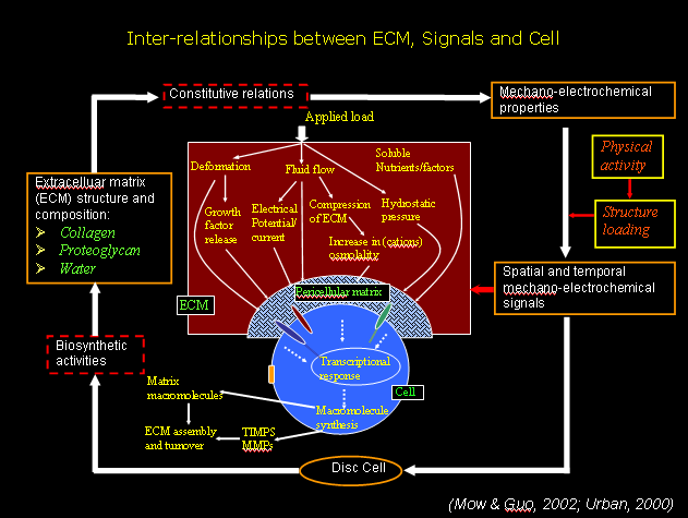
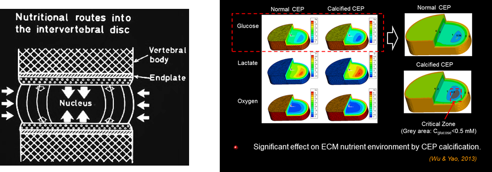
Low back pain is a major socio-economic concern in the United States, affecting more than 70% of individuals at some point in their lifetime. Although the exact cause is unclear, degeneration of the intervertebral disc (IVD) has been implicated as a possible primary etiological factor. Since IVDs are the largest avascular tissue in the human body, transport of essential nutrients is crucial to maintain disc health. Deviation from physiological nutrient levels due to abnormal mechanical loading and age is suggested to be one of the main mechanisms for disc degeneration. The goal of this research is to measure transport properties and cell metabolic rates of human IVDs, and to develop a new mechano-electrochemical transport theory (mixture theory based) and finite element model (FEM) for investigating the transport of fluids and solutes and cell metabolic activities in vivo under mechanical stress in normal and degenerated human IVDs. The advance in theory, numerical tools, data on tissue properties and cell metabolic rates, and measuring techniques will have a significant impact on understanding etiology of disc degeneration as well as on developing new strategies for tissue regeneration.
3. Ocular Biomechanics and Biotransport
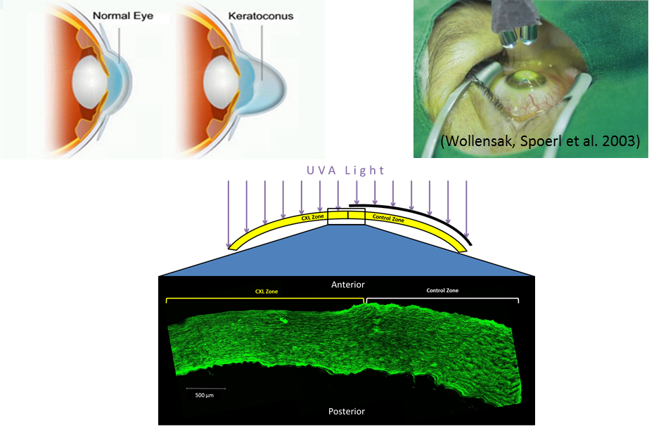
Approximately 1 in 2000 people suffer from keratoconus, a disease characterized by a weakening and protrusion of the cornea that leads to astigmatism. 21% of keratoconus patients have a progressive form that eventually requires a corneal transplant. A decade ago, Wollensak et al. reported a new treatment for keratoconus that strengthens the cornea through crosslinking with riboflavin and UVA light. Since it was first presented clinically a decade ago, various protocols for riboflavin delivery and UVA irradiation have been developed, with varied results. The goal of this research is to develop sensitive tests and comprehensive finite element models to compare and predict the outcomes of these protocols, as well as to understand the crosslinking mechanism.
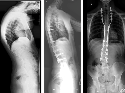
4. Biomechanics of Early Onset Scoliosis
Early onset scoliosis is a condition with multiple etiologies that has an incidence rate of 1-2 out of 10,000. Severe cases are treated with spinal implants. Current methods of fixation have been reasonably successful; but when kyphosis is also present, failure is more common. The broad, long-term objective of this study is to (1) determine the strength of currently used instrumentation to kyphotic force load, (2) understand the mechanism of implant failure, and (3) develop a new method to predict implant design success based on thoracic fixation.